Home
/ Bone Cross Section Diagram Labeled - Structure Of Long Bone Animal Systems _ Cross section through the thalamus:
Bone Cross Section Diagram Labeled - Structure Of Long Bone Animal Systems _ Cross section through the thalamus:
Bone Cross Section Diagram Labeled - Structure Of Long Bone Animal Systems _ Cross section through the thalamus:. Related posts of bone cross section labeled. The diaphysis and the epiphysis. Indicated are haversian canal with blood and lymphatic vessels, a nerve, and loose. The outside of a bone is covered in a thin layer of dense irregular connective tissue called the periosteum. Bodytomy provides a labeled diagram of the haversian system to help you the terms 'haversian system' or 'osteon' refer to the basic.
Cross section of a bone. Compact bone is the denser, stronger of the two types of bone tissue ( (figure) ). Smartdraw includes 1000s of professional healthcare and anatomy chart templates that you can modify and make your own. Jump to navigation jump to search. The compact bone is made up of osteon.
Cartilage Bone Ossification The Histology Guide from www.histology.leeds.ac.uk Cross section of a bone. Personalize it with photos & text or purchase as is! A long bone has two parts: Looking at a bone in cross section, there are several distinct layered regions that make up a bone. Cookies allow us to analyze and store information such as the characteristics of your device as well as certain personal data (e.g., ip addresses, navigation, usage or geolocation data, unique identifiers). Bone decalcification is the removal of the mineral component using an acid, leaving the bone soft and easy to cut. The diaphysis is the tubular shaft that runs between the proximal and distal ends of the bone. The diaphysis and the epiphysis.
The diagram above shows a transverse view.
Bone matrix and cells bone matrix osseous tissue is a connective tissue and like all connective tissues contains relatively few cells and large amounts of extracellular matrix. At a symphysis, the bones are joined by fibrocartilage. Related posts of cross section of a long bone bone test anatomy and physiology. Personalize it with photos & text or purchase as is! The diaphysis and the epiphysis. Download 706 bone cross medical section stock illustrations, vectors & clipart for free or amazingly low rates! The diagram above shows a transverse view. It can be found under the periosteum and in the diaphyses of long bones, where it provides support and protection. Bones in your body names. Which labeled structure in the given image is a fascicle? Cross section of a long bone. Related posts of bone cross section labeled. The point of attachment on the more movable bone in the illustration is called the _____ of the muscle.
Find the perfect bone cross section stock photos and editorial news pictures from getty images. Red marrow fills the spaces in some bones. Human skeleton anatomy human body anatomy human anatomy and physiology muscle anatomy hand bone anatomy anatomy bones the human body anatomy images anatomy practice. New users enjoy 60% off. The structure of a long bone allows for the best visualization of all of the parts of a bone ( figure 6.7 ).
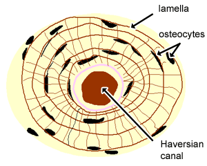
Mastering A P Chapter 6 Bones And Skeletal Tissues Flashcards Quizlet from o.quizlet.com Download scientific diagram | schematic of osteon. I don't like way you've shown the cartilage. It seems confusing and misleading. The diaphysis is the tubular shaft that runs between the proximal and distal ends of the bone. Diagram orienting yourself within such a cross section is easy. Free online quiz compact bone microscope slide labeled. Shop bone cross section diagram label created by chartsanddiagrams. The central haversian canal, and horizontal canals (perforating/ volkmann's) canals contain blood vessels and nerves from the periosteum.
The star of the show (brain) is easily recognizable because it appears highly convoluted, full of ridges (gyri) and indentations (sulci).the paired thalami appear as two circular masses in the midline, forming the walls of the third ventricle.the neurocranium appears as a meshwork (trabecular.
Human skeleton anatomy human body anatomy human anatomy and physiology muscle anatomy hand bone anatomy anatomy bones the human body anatomy images anatomy practice. From which part of the sarcomere is the given cross section taken? A cross section of a human long bone. Damage in even one part can hinder the functioning of the knee. Related posts of cross section of human bone diagram bone in arm pictures. Download 706 bone cross medical section stock illustrations, vectors & clipart for free or amazingly low rates! This photo shows a cross section through bone. There are two ways to study bone histology. Shop bone cross section diagram label created by chartsanddiagrams. Fixed slide cross section of muscle tissue, 100x microscope view. Find the perfect bone cross section stock photos and editorial news pictures from getty images. Looking at a bone in cross section, there are several distinct layered regions that make up a bone. New users enjoy 60% off.
Indicated are haversian canal with blood and lymphatic vessels, a nerve, and loose. There are two ways to study bone histology. Shop bone cross section diagram label created by chartsanddiagrams. Cross section of human bone diagram 12 photos of the cross section of human bone diagram cross section diagram of human bone, bone, cross section diagram of human bone. Cookies allow us to analyze and store information such as the characteristics of your device as well as certain personal data (e.g., ip addresses, navigation, usage or geolocation data, unique identifiers).
Muscular And Skeletal Systems from www2.estrellamountain.edu Download 706 bone cross medical section stock illustrations, vectors & clipart for free or amazingly low rates! Browse 4,294 bone cross section stock photos and images available, or search for human bone cross section to find more great stock photos and pictures. Storage of fat for a ready energy source for active muscles. Anatomy of a flat bone. Bodytomy provides a labeled diagram of the haversian system to help you the terms 'haversian system' or 'osteon' refer to the basic. Damage in even one part can hinder the functioning of the knee. The muscle acts as the effort force; Imaios and selected third parties, use cookies or similar technologies, in particular for audience measurement.
Cookies allow us to analyze and store information such as the characteristics of your device as well as certain personal data (e.g., ip addresses, navigation, usage or geolocation data, unique identifiers).
Browse 4,294 bone cross section stock photos and images available, or search for human bone cross section to find more great stock photos and pictures. Bone test anatomy and physiology 12 photos of the bone test anatomy and physiology anatomy and physiology bone lab test, anatomy and physiology bone markings test, anatomy and physiology bone practical test, anatomy and physiology bone tissue test, anatomy and physiology test on bone tissue, bone, anatomy and. Damage in even one part can hinder the functioning of the knee. The structure of a long bone allows for the best visualization of all of the parts of a bone ( figure 6.7 ). Cross section of human bone diagram 12 photos of the cross section of human bone diagram cross section diagram of human bone, bone, cross section diagram of human bone. A long bone has two parts: The central haversian canal, and horizontal canals (perforating/ volkmann's) canals contain blood vessels and nerves from the periosteum. Jump to navigation jump to search. The star of the show (brain) is easily recognizable because it appears highly convoluted, full of ridges (gyri) and indentations (sulci).the paired thalami appear as two circular masses in the midline, forming the walls of the third ventricle.the neurocranium appears as a meshwork (trabecular. The osteocytes are arranged in concentric rings of bone matrix called lamellae (little plates), and their processes run in interconnecting canaliculi. Diagram orienting yourself within such a cross section is easy. Human skeleton anatomy human body anatomy human anatomy and physiology muscle anatomy hand bone anatomy anatomy bones the human body anatomy images anatomy practice. At a symphysis, the bones are joined by fibrocartilage.
The structure of a long bone allows for the best visualization of all of the parts of a bone ( figure 67 ) bone cross section. Bones in your body names.

